
Sample report · Cat X-ray
Sample report: Luna's chest X-ray and an enlarged heart
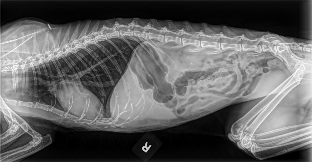
A real chest-and-abdomen X-ray from a domestic shorthair cat whose vet had heard a gallop rhythm, exactly as the owner saw it on Read Your Pet's Lab — only the name has been changed. It shows how a report handles a finding that matters without being alarming, and how it separates what the film shows from what it cannot answer.

Pet
Luna
adult spayed female domestic shorthair, 3.5 kg
What the owner told us
“Gallop rhythm, weight loss, pancreatitis, not eating well, urine specific gravity 1.020.”
Right lateral view of the chest and front of the abdomen
X-ray · Veterinary imaging report
X-ray Analysis
Explanation for Pet Owners
Luna’s heart silhouette appears enlarged and should be evaluated
Luna’s single side-view X-ray shows an apparently enlarged heart silhouette, without obvious fluid around the lungs or a clear pattern of lung congestion on this image. Because a gallop rhythm has been reported, her veterinarian may recommend a heart ultrasound and additional chest views. The abdominal portion does not show an obvious intestinal blockage, although this limited view cannot assess every cause of weight loss or poor appetite.
Key Points
- The heart appears larger than expected for a cat, which should be discussed with Luna’s veterinarian.
- There is no obvious lung edema or pleural fluid on this single view.
- The visible intestines contain some gas, but there is no clear radiographic sign of an obstruction.
- Several small metallic densities over the chest are most consistent with prior surgical material or external artifacts.
Findings marked on the X-ray
In a real report, tapping a finding highlights it on the image.
Recommended Next Steps
- Arrange prompt veterinary review, particularly because Luna has a reported gallop rhythm, weight loss, and reduced appetite.
- Ask whether a complete three-view chest study and echocardiogram (heart ultrasound) are appropriate.
- Continue the veterinarian’s evaluation of pancreatitis and other causes of weight loss; abdominal ultrasound may be more informative than this X-ray for those concerns.
Questions You May Want to Ask Your Vet
In a real report, tapping a question sends it to the follow-up chat.
Clinical Summary
Domestic Shorthair cat, single right lateral radiograph including the thorax and cranial-to-mid abdomen. The image appears to be a clean digital radiograph without obvious monitor glare, moire, or screen artifacts. Interpretation is limited because only one projection is provided and the caudal abdomen is incompletely included.
Findings
- Cardiac silhouette is subjectively enlarged, with generalized enlargement suspected; exact vertebral heart size is not reliable on this single lateral view.
- Pulmonary vessels are not obviously enlarged, and there is no convincing pulmonary alveolar edema identified on this view.
- No pleural effusion or pneumothorax is evident.
- Mild diffuse bronchial/interstitial pulmonary opacity may be present but is not definitive on this image.
- Visible small intestinal and colonic gas is present without clear segmental severe dilation, plication, or obstructive pattern.
- Multiple small linear metallic opacities project over the ventral thorax, most compatible with postsurgical material or overlying artifacts.
- No obvious radiopaque gastrointestinal foreign body or aggressive osseous lesion is identified within the included field.
Impression
- Suspected cardiomegaly without radiographic evidence of congestive pulmonary edema on this single view.
- The cardiac finding is clinically relevant given the reported gallop rhythm; echocardiographic assessment is recommended.
- No obvious radiographic gastrointestinal obstruction in the included abdominal region.
Recommendations
- Obtain orthogonal thoracic views, preferably a complete three-view thoracic series, if clinically indicated.
- Recommend echocardiography and consideration of blood pressure measurement and ECG based on the reported gallop rhythm.
- Consider abdominal ultrasound and laboratory correlation for persistent weight loss, anorexia, or suspected pancreatitis.
AI Disclaimer
This is an AI-generated educational explanation, not a diagnosis, and it does not replace examination by a veterinarian. A single X-ray view may not capture all abnormalities.
Important Notes
- Seek urgent veterinary care if Luna develops rapid or difficult breathing, open-mouth breathing, collapse, marked weakness, or blue/pale gums.
- An enlarged heart on one radiograph does not establish the specific cause; heart ultrasound is usually needed for characterization.
- The reported gallop rhythm, weight loss, and poor appetite should be evaluated by Luna’s veterinarian even if breathing currently appears normal.
What you're looking at
One amber point, three green ones
The report leads with the one finding that matters — a heart that looks larger than expected for a cat — and then says clearly what it does not see: no fluid around the lungs, no obvious lung congestion, no sign of a blockage. That balance is deliberate. An enlarged heart silhouette is a reason to book an echocardiogram, not a reason to panic tonight.
The film can't answer the pancreatitis question
X-rays are good at bone, air and the outline of organs. They are poor at the pancreas and at explaining weight loss. The report says so, and points the owner towards abdominal ultrasound and bloodwork — which is exactly what the vet would say.
Those tiny bright dots
The small metallic dots over the chest look dramatic to an owner. The report reads them as leftover surgical material or something lying on the table — common, and nothing to do with the heart.
Your words go in, better questions come out
The owner typed five clinical words from the consult. The report used them to shape its reading and returned three precise questions about echocardiography, blood pressure and ultrasound that the owner could ask at the next visit.
Get your own cat's X-ray explained
Upload the image your vet gave you — a JPEG, PNG or DICOM file, or a clear photo of the screen — and add anything the vet said. You get the same plain-language walk-through for your cat within minutes. Your first analyses are free, no credit card.
Free to start · no credit card · educational, not a diagnosis
More sample reports
Different pets, different questions — the same plain-language approach.

Read Your Pet's Lab is an educational tool. It explains what results mean and helps you prepare for your vet visit — it does not diagnose, and it never replaces the veterinarian who examined your pet.