
Sample report · Dog X-ray
Sample report: did Max swallow something?
This is a real abdominal X-ray series from a dog who had been vomiting for three days. Two views were taken, and they tell slightly different stories. That is normal, and it is the most useful thing this sample can show you.

Pet
Max
3-year-old neutered male Australian Shepherd
What the owner told us
“Vomiting for three days. Our vet is worried he swallowed something and mentioned a dilated duodenum.”
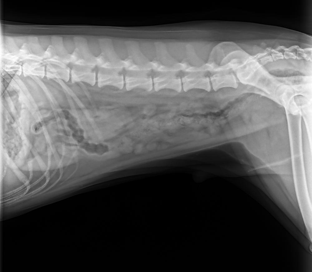
View 1 · right lateral (side) view of the abdomen
X-ray · Veterinary imaging report
X-ray Analysis
Explanation for Pet Owners
Stomach contents need prompt veterinary follow-up
Max’s X-ray shows abnormal material and gas/fluid within the stomach region, and the nearby intestinal tract may be somewhat distended. This can occur with ordinary stomach contents, but a swallowed object cannot be confirmed or excluded from this single view. Because Max has been vomiting for three days, he should be evaluated promptly by his veterinarian.
Key Points
- There is material in Max’s stomach that could represent ingesta or a swallowed foreign object.
- Some gas-filled bowel is visible near the stomach, but one view cannot reliably determine whether an obstruction is present.
- No obvious free abdominal gas is seen on this view, which is reassuring against a clearly perforated bowel.
- Many swallowed objects, including fabric or plastic, may not be visible directly on X-rays.
Findings marked on the X-ray
In a real report, tapping a finding highlights it on the image.
Recommended Next Steps
- Contact Max’s veterinarian promptly today because of the three-day vomiting history.
- Ask whether additional abdominal views and an abdominal ultrasound are recommended to evaluate for obstruction or a gastric foreign body.
- Do not give Max food, medications, or induce vomiting unless his veterinarian specifically instructs you.
Questions You May Want to Ask Your Vet
In a real report, tapping a question sends it to the follow-up chat.
Clinical Summary
Clean digital-appearing right lateral abdominal radiograph of a 3-year-old Australian Shepherd. The abdomen is included in a single lateral projection; no obvious image-capture degradation is present. Interpretation is limited because an orthogonal view is not available.
Findings
- Moderate mixed gas, fluid, and soft-tissue opacity within the gastric lumen.
- Small irregular mineral-opaque material projects over the gastric region and is suspicious for mineral-opaque gastric foreign material, although ingesta cannot be excluded.
- Mild focal gas distention of proximal small intestinal segments is suspected; the degree and significance of duodenal distention cannot be confidently assessed on this single view.
- No definitive generalized small intestinal obstructive pattern is identified on the provided projection.
- No obvious pneumoperitoneum is seen on this view; serosal detail is generally maintained.
Impression
- Gastric contents include mineral-opaque material suspicious for, but not diagnostic of, a gastric foreign body.
- Possible mild proximal small intestinal distention; mechanical obstruction cannot be excluded, particularly given the reported persistent vomiting.
- Further imaging is warranted for clarification.
Recommendations
- Obtain orthogonal abdominal radiographs, including a ventrodorsal or dorsoventral view.
- Recommend abdominal ultrasound to assess gastric emptying, the pylorus and duodenum, bowel wall, and any radiolucent foreign material.
- Urgent veterinary assessment is appropriate if vomiting persists or if Max becomes painful, weak, bloated, or unable to keep water down.
AI Disclaimer
This is an AI-generated educational explanation, not a diagnosis, and it does not replace examination by a veterinarian. A single X-ray view may not capture every abnormality.
Important Notes
- Persistent vomiting with possible obstruction can become urgent; contact Max’s veterinarian promptly.
- Seek emergency care for marked abdominal pain or swelling, repeated unproductive retching, collapse, severe weakness, or inability to keep water down.
- Do not induce vomiting unless directed by a veterinarian.
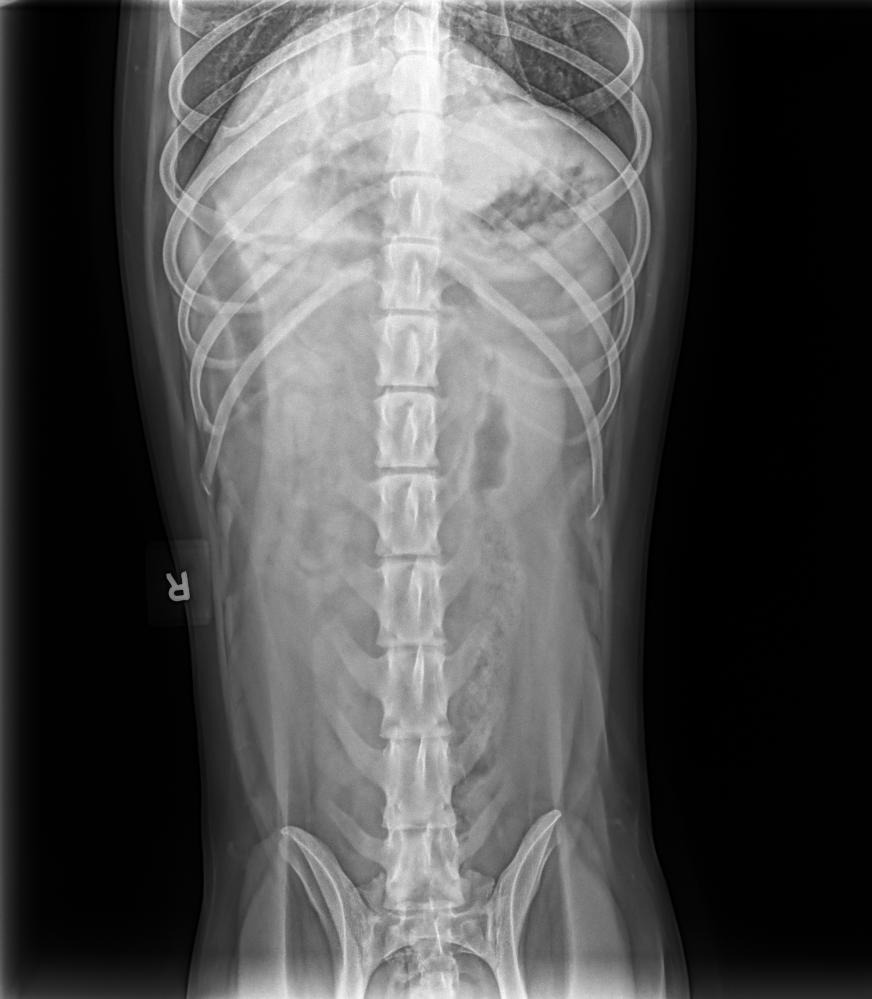
View 2 · ventrodorsal (top-down) view of the abdomen
X-ray · Veterinary imaging report
X-ray Analysis
Explanation for Pet Owners
No obvious blockage or metal-like foreign object is visible on this abdominal view
Max’s single abdominal X-ray does not show a clearly visible radiopaque foreign object or an obvious pattern of intestinal blockage. There is a small amount of gas in the stomach and proximal intestinal region, which can occur normally, but one view cannot rule out a soft or radiolucent object or an early obstruction.
Key Points
- No clearly visible mineral or metal-like foreign object is identified.
- The stomach and intestines are not obviously enlarged in an obstructive pattern on this view.
- A small amount of gas near the proximal duodenum is visible but is not clearly abnormal by itself.
- Many foreign objects do not show on X-rays, so a foreign body cannot be completely excluded.
Recommended Next Steps
- Have Max’s veterinarian compare this with a lateral abdominal view and his examination findings.
- If vomiting, abdominal pain, poor appetite, or ongoing concern persists, abdominal ultrasound may be helpful.
- Seek prompt veterinary care for repeated vomiting, unproductive retching, a painful or swollen abdomen, marked lethargy, or inability to keep water down.
Questions You May Want to Ask Your Vet
In a real report, tapping a question sends it to the follow-up chat.
Clinical Summary
Max is a 3-year-old neutered male Australian Shepherd. This is a clean digital ventrodorsal abdominal radiograph, without evident monitor-recapture or significant image degradation. It is a single screening projection obtained in the context of concern for a gastric foreign body and gas in the duodenum.
Findings
- The stomach contains a small amount of gas and soft-tissue/fluid opacity but is not markedly distended.
- A small amount of gas is present in the expected proximal duodenal region; there is no convincing focal duodenal dilation on this view.
- Small intestinal loops are not diffusely or markedly distended, and no clear obstructive gas pattern is identified.
- No discrete radiopaque foreign body is definitively visible.
- Serosal detail is adequate on this view, with no evidence of free intraperitoneal gas.
- The included osseous structures have no acute abnormality identified on this screening image.
Impression
- No radiographic evidence of a high-grade gastrointestinal obstruction on this single ventrodorsal view.
- Mild proximal gastrointestinal gas, including gas in the expected duodenal region, is nonspecific and may be within physiologic limits.
- A radiolucent foreign body or early/partial obstruction cannot be excluded radiographically.
Recommendations
- Obtain a right or left lateral abdominal projection, ideally with additional orthogonal views if clinically indicated.
- Consider abdominal ultrasound if clinical signs persist or suspicion for a non-radiopaque foreign body remains.
- Serial abdominal radiographs may help assess progression when obstruction is a concern.
AI Disclaimer
This is an AI-generated educational explanation, not a diagnosis, and it does not replace examination by a veterinarian. A single X-ray view may not capture every abnormality.
Important Notes
- Because soft objects may not be visible on X-rays, persistent clinical concern should be assessed by Max’s veterinarian.
- Do not induce vomiting or give laxatives unless specifically instructed by a veterinarian.
- Seek urgent care for repeated vomiting, retching, abdominal distension or pain, severe lethargy, or inability to keep water down.
What you're looking at
Two views, two readings — and that's the point
The side view shows small mineral-dense flecks over the stomach and raises the possibility of a foreign object. The top-down view, taken a minute later, shows no dense object at all. Neither is wrong: X-rays are shadows, and a small object can hide behind stomach contents in one direction and not the other. This is exactly why vets take at least two views, and why "nothing obvious" on one film is not the same as "nothing there".
The colours tell you how to feel about each point
Amber points are the ones worth a conversation; green points are reassuring. Here the report is careful: it flags what it sees, says what it cannot see, and tells the owner what would turn this into an emergency.
Soft objects don't show up
Fabric, plastic, string and most toys are radiolucent — they let X-rays through and leave no shadow. If a dog has been vomiting for days, a clean-looking X-ray is reassuring but not conclusive, which is why ultrasound is the next step the report suggests.
The questions are the part you take to the appointment
Every report ends with three questions written for this pet and this film. Owners tell us this is the part they actually use: it turns an anxious wait into a short, focused conversation with the vet.
Get your own dog's X-ray explained
Upload the image your vet gave you — a JPEG, PNG or DICOM file, or a clear photo of the screen — and you get the same plain-language walk-through for your dog within minutes. Your first analyses are free, no credit card.
Free to start · no credit card · educational, not a diagnosis
More sample reports
Different pets, different questions — the same plain-language approach.

Read Your Pet's Lab is an educational tool. It explains what results mean and helps you prepare for your vet visit — it does not diagnose, and it never replaces the veterinarian who examined your pet.